IT IS OFTEN said that the eyes are the windows to the soul. Researchers hope that they might also be a window to the brain. Scientists wonder if eye scans could provide information about a wide range of conditions, including ADHD, Alzheimer’s disease, autism, schizophrenia and traumatic brain injury. They are already used to detect predispositions to physical-health problems like high blood pressure and diabetes. On August 21st researchers at Moorfields Eye Hospital and University College London’s Institute of Ophthalmology published a paper in which they said they had identified markers of Parkinson’s disease in the eye seven years before it would have been apparent using existing tests. How?
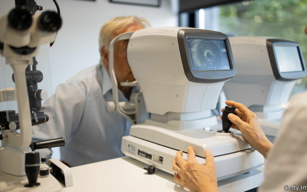
Changes in the eye, particularly the retina, sometimes appear to reflect changes in the brain. The retina is like a piece of wet tissue paper at the back of the eye that contains light-sensitive nerve cells in many distinct layers. It grows from the same tissue as the brain during embryonic development and is connected to the brain by the optic nerve. It thus shares many of the brain’s characteristics. If a relationship between brain and retina were proved, it could be extremely useful: brains are difficult to study while their owners are alive. Eyes, on the other hand, are easy to scan in detail with equipment found in the average high-street optician’s office. The technique in question is optical coherence tomography (OCT), a non-intrusive 3-D scan that works by bouncing light waves across the eye, and taking pictures of the retina and each of its layers, which are then mapped and measured.
Scientists have long suspected that the thinning of the retina may be an indicator of Parkinson’s. In the new paper, published in Neurology, researchers have shown this relationship in a fairly comprehensive way. First they used machine learning to analyse OCT scans of more than 150,000 eye-hospital patients over the age of 40. One layer of the retina in particular, the ganglion cell-inner plexiform layer, which contains the nuclei of nerve cells, was found to be thinner in patients who went on to develop Parkinson’s. (Changes were also found in another layer that contains dopamine-producing neurons in the eye.) The relationship was tested in a second cohort of about 67,000 patients from a medical database, confirming the link. The technique is not yet accurate enough to predict whether a person will develop Parkinson’s: some people with the marker won’t develop Parkinson’s and some people with Parkinson’s don’t have the marker. But it could be a simple pre-screening tool, and could easily be integrated into routine eye tests.
The hunt for ocular “biomarkers” is a promising area of research. Pearse Keane, one of the authors of the recent Parkinson’s study, says that scientists have known for more than a century that signs of diseases in the body can be detected with eye tests. Researchers hope that the proliferation of OCT machines and advances in artificial intelligence will supercharge their efforts. Ocular markers might one day help with efforts to prevent or slow the onset of degenerative diseases and even identify people suitable for trials of new drugs. Some envisage a day when they will guide personalised treatments. At the very least, this research is worth keeping an eye on. ■


